Cellular/molecular imaging of biomarkers and therapies in brain disease models
In many brain disorders – including stroke, dementia, multiple sclerosis and epilepsy – critical pathophysiological processes occur at the neurovascular level. These include cerebral blood flow abnormalities, blood-brain barrier disruption, neuroinflammation and cerebrovascular remodeling.
Research participants
EL Blezer, RM Dijkhuizen & G van Tilborg
Contrast-enhanced MRI
With contrast-enhanced MRI of passage or leakage of intravascular paramagnetic contrast agent, we can quantify perfusion deficits or blood-brain barrier permeability, respectively. Moreover, recent advances in contrast agent design – allowing agents to be long-circulating, cell-loadable and/or targeted – have enabled measurement of tissue vascularity, cellular infiltration and endothelial markers with MRI. This requires interdisciplinary cross-talk, for which we have teamed up with (inter)national experts in nanotechnology, cellular and molecular biology, and pharmacology.
Gradient- and spin-echo MRI acquisition
Cerebrovascular indices, such as cerebral blood volume, microvascular density and vessel size index, can be quantified from combined gradient- and spin-echo MRI acquisition after injection of a superparamagnetic blood pool agent. Vascular steady state contrast-induced signal changes, depending on morphological properties of the vascular network, can thereby expose various features of the vasculature associated with brain pathology or plasticity.
Cell labeling with superparamagnetic contrast agents
Cell labeling with superparamagnetic contrast agents – under in vivo or in vitro conditions – allows MRI-based detection of labeled cells. This approach can be employed to measure infiltration of leukocytes, like monocytes and T-lymphocytes, which can play a critical role in brain pathophysiology, but may also be of use to monitor delivery and fate of stem cells in cell-based therapies.
Targeted contrast agents
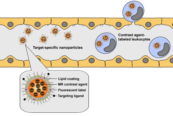
MRI of molecular entities is feasible with targeted contrast agents. Targeting can be accomplished by conjugation of a specific ligand – e.g. an antibody – to a contrast agent complex. We have shown that endothelial markers, such as cell adhesion molecules involved in binding and infiltration of circulating leukocytes, can be detected following intravascular injection of selectively binding contrast agents.

Schematic overview of the concept of cellular and molecular MRI of neuroinflammation. Target-specific contrast agents allow detection of upregulated endothelial markers, such as cell adhesion molecules, while infiltrating leukocytes can be detected after specific cell labeling with contrast agents. Multimodal lipid-based nanoparticles with incorporated MR contrast agent and fluorescent label can be designed for combined MRI and optical imaging of cells or molecular markers. Courtesy of Geralda van Tilborg.
Contrast-enhanced MRI
Contrast-enhanced MRI methods provide powerful means to assess (changes in) hemodynamic indices, vascular parameters and inflammatory markers, with the potential of identifying critical biomarkers and therapeutic targets. These approaches thereby broaden the already impressive arsenal of MRI methods for experimental and (pre)clinical assessment of brain diseases and therapies.

T2*-weighted MR images of a mouse brain slice before and 1 h after injection of micron-sized particles of iron oxide coated with antibodies against intercellular adhesion molecule-1 (αICAM-1-MPIO), at 24 h after ischemic stroke. The lesion is characterized by edema-associated signal increase in pre- and post-contrast images, while a vascular pattern of hypointensities, reflecting neurovascular inflammation, appeared after αICAM-1-MPIO injection. Courtesy of Lisette Deddens.
