Image guided intratumoral treatment of malignancies
Cancer is among the leading causes of death worldwide accounting for 8.2 million deaths and 14.1 million patients were diagnosed with cancer in 2012. Depending on the patient’s medical status and stage of disease, systemic cytotoxic chemotherapy and/or surgery are the mainstay for cancer therapy. However, for some cancers such as brain, lung, pancreas and liver, the treatment options are often only palliative with a dismal prognosis of only 1 year. These figures call for a major transformation of the therapeutic management of cancer.

Research participants
Rob Bakker, MA van den Bosch, Robert van Es (Oral and Maxillofacial Surgery), MG Lam, JF Nijsen & Bas van Nimwegen (veterinary medicine)
Radioembolisation uitklapper, klik om te openen
In recent years more personalised local treatment options are emerging to improve patient outcomes. One of these promising therapies is radioembolisation where radioactive microspheres are intra-arterially injected through imaging-guided catheterization. This treatment is currently used for selected patients with liver tumors resulting in good response rates, minor side-effects and good quality of life. The Holmium research group has pioneered the preclinical and clinical development of radioactive holmium microspheres (166Ho-MS) as a new (image-guided) treatment approach for patients with liver cancer. To advance the use of 166Ho-MS for malignancies in other organs, a direct injection strategy is currently under investigation.
Direct intratumoral injection of β-emitting radioactive holmium microspheres (microbrachytherapy) has been shown to result in a high tumor absorbed dose, and could be an alternative for current treatments. The shallow tissue penetration of β-radiation permits very high local tissue radioactive dose while the surrounding healthy tissue is spared. Therefore, biocompatible microspheres loaded with holmium-166 (166Ho; Eβ,max=1.84 MeV; T1/2 = 26.8 h) were developed for this intratumoral approach. The Holmium microspheres(166Ho-MS) can be visualized and quantified in vivo by MRI, CT and SPECT, enabling the possibility of real-time feedback of microsphere spatial and dose distribution. This concept needs to be thoroughly investigated in a preclinical setting.
The aim of this project is to develop an effective minimally invasive therapeutic strategy that is based on an innovative method of image guided local application of radioactive microspheres within the tumor boundary. The research that is performed fits into a bench-to-bedside approach of translational research. Therefore, there is a close cooperation with companies and patients.
Animal patients uitklapper, klik om te openen

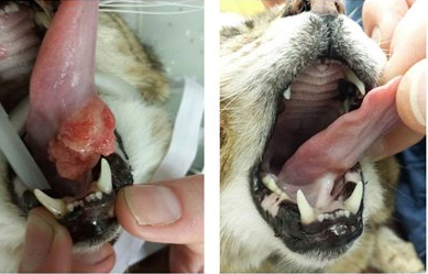
Cat with a malignant tumor on the tongue (left) and result after treatment with radioactive holmium microspheres (right).
The Holmium Research Group and the Utrecht University (UU) Animal Cancer Center have a close collaboration on the holmium microsphere technique. The Animal Cancer Center attracts many referral veterinary patients for diagnostic and treatment procedures. During the last 5 years over 35 veterinary patients (cats and dogs) with advanced stages of tumors that were beyond surgical resection were treated by injection of 166Ho-MS directly into the tumors . In general, all responders markedly improved clinically. Toxicity was absent in most animals and a partial or complete response was achieved which frequently led to down staging enabling subsequent surgical resection. It is observed that intratumoral administration of 166Ho-MS, is feasible and effective in veterinary patients and is well-tolerated and not associated with (serious) adverse side effects.
