Back
Myocardial fibrosis detection without a contrast agent
Our aim is to detect myocardial fibrosis quantitatively after different cardiomyopathies. Preferably this imaging is done without the use of a contrast agent.
T. Leiner / J.W. van Oorschot / J.J. Zwanenburg
T1ρ-mapping technique
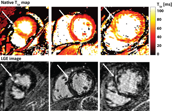
We have developed a cardiac T1ρ-mapping technique which is used to detect fibrosis in the heart, and have shown the first clinical results for the detection of chronic myocardial infarction with the use of native T1ρ-mapping. Furthermore we are studying other MRI contrasts such as T1-, T2- and ECV-mapping.
Figure 1: Short axis T1ρ-maps with corresponding LGE images in 3 different patients. Arrows indicate the infarcted area.

