Kidney tumors in children: specific diagnosis with MRI

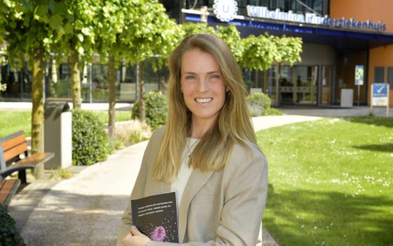
Kidney tumors are rare amongst children, and their treatment can be intensive. Justine van der Beek researched how different types of kidney tumors can be distinguished using MRI scans. This technique could help provide the best treatment for each child from the early diagnosis stage. "A faster personalized treatment contributes to a better chance of recovery and quality of life " says Justine, who works at the Princess Máxima Center and UMC Utrecht. She earned her PhD on May 28.
In the Netherlands, about 35 children are diagnosed with a kidney tumor each year. Most often, this is a Wilms tumor, which primarily occurs in children under five. Due to international collaboration and research, significant progress has been made, and 9 out of 10 children recover.
In Europe, children first receive chemotherapy before the tumor is removed through surgery. "Because it is so likely to be a Wilms tumor, all children with kidney cancer receive the standard treatment for Wilms tumors, which is 4 to 6 weeks of chemotherapy," Justine explains. Additionally, Wilms tumors often consist of multiple subtypes of tumor cells. "The goal of chemotherapy is to shrink the tumor before surgery, which makes the operation easier," Justine explains. "However, we see that this treatment does not always have the desired effect on certain (sub)types, meaning children sometimes undergo intensive therapies without shrinking the tumor."
Distinguishing tumors
Currently, distinguishing Wilms tumors from non-Wilms tumors at the time of diagnosis can only be done by taking a small piece of the tumor (biopsy) for examination under a microscope. A biopsy is taken under anesthesia using ultrasound guidance through the skin. Pediatric oncologists in Europe only perform this when they suspect it is not a Wilms tumor, as a biopsy carries risks. A less invasive way to determine the type of kidney tumor would be beneficial.
Justine investigated how MRI-scans can help distinguish Wilms tumors from non-Wilms tumors, and also looked at distinguishing subtypes of Wilms tumors. Since all children with a kidney tumor routinely receive an MRI-scan at diagnosis and after chemotherapy, this method does not add any extra burden to the child. "In this way, we hope to be able to offer truly tailored treatments from the very beginning, determining the exact diagnosis and the best treatment for each patient," says Justine.
Diagnosing with MRI
In her research, Justine used a special MRI technique called diffusion-weighted imaging (DWI). With DWI, Justine measured how quickly water molecules move in the tissue of the kidneys and tumors. "Tumor cells are more densely packed than normal cells, causing water molecules to move more slowly. The speed of water molecule movement also appears to differ per subtype. By looking at this cell density, we can distinguish different tumor types," Justine explains.
To precisely determine the (sub)type of tumor cells, Justine and her colleagues used a personalized 'cutting guide'. This is a 3D-printed mold that fits the kidney tumor exactly. "This allowed us to examine the subtypes under the microscope and directly compare them to the MRI scan. We made DWI measurements linked to specific subtypes of kidney tumors", Justine says. The values obtained help to distinguish kidney tumors in children, along with other characteristics of the different types of kidney tumors.
International collaboration
Justine conducted her PhD research in collaboration with UMC Utrecht, Wilhelmina Children's Hospital (WKZ), and Princess Máxima Center (PMC) under the guidance of Prof. Jeroen Hendrikse, Prof. Marry van den Heuvel-Eibrink, Prof. Ronald de Krijger, and Dr. Annemieke Littooij. International collaborations are also important in this research. "Fortunately, cancer in children is rare, but because it is so rare, we can achieve a lot by joining forces nationally and internationally," Justine explains. "At the Princess Máxima Center, we see how far we can come by working together, especially as we can focus on very specific methods for certain tumor types."
Next steps
Justine is optimistic that with more research and collaboration between specializations, these new MRI-based methods will lead to treatments specifically tailored to each child's tumor type. This way, every child can receive the best possible care in the future. Justine: "I truly believe that with this dissertation, we are making a big leap forward and have laid the foundation for future research."
The results of the feasibility study, made possible by funding from KiKa, were previously published in Pediatric Oncology. The results of the follow-up study will be published soon. Biopsies and scans from 54 children treated at the Máxima have been examined for this study.
