Single cell RNA sequensing uncovers new mechanisms of heart disease

Hypertrophic cardiomyopathy is a heart disease that leads to a stressed, swollen heart muscle. Due to a poor understanding of underlying mechanisms, effective clinical treatments are not available. Patients receive generic heart medication and sometimes need open-heart surgery to remove excess tissue. Researchers at the Hubrecht Institute have now successfully applied a new revolutionary technology (scRNA-seq) to uncover underlying disease mechanisms, including specifically those causing the swelling. The extensive “big data” set is a treasure trove of novel observations that give insight in hypertrophic cardiomyopathy and potential new therapeutic venues. The results from this study, done by researchers in the group of Eva van Rooij, were published in the journal Cell Reports on the 10th of May.
The heart needs to pump every minute of every day. In patients with hypertrophic cardiomyopathy (HCM), this pumping function is impaired because of a defect in one of the molecules that perform the pulling motion. This leads to a stress response within the muscle cells, and swelling of the heart muscle to compensate for lost function. As a consequence, patients can experience typical heart disease symptoms like shortness of breath, chest pain and aberrant heart rhythm (arrhythmia). Up until today, development of HCM therapy is hindered by a lack of understanding of these phenomena.
Cogwheels
Surgery on HCM patients to remove excess heart tissue that hinders blood flow offers a unique opportunity to researchers, because they can use the removed tissue to study the disease. Hubrecht Institute researchers have now applied the novel single cell RNA sequencing (scRNA-seq) technology on this tissue to unravel the origins of HCM. One of the researchers, Martijn Wehrens, explains: “The human DNA contains approximately 30,000 genes, effectively a catalogue of 30,000 types of cogwheels that each have a role in making our bodies work. Typically, research focuses on a handful of genes, that have been identified as important after years of research. scRNA-seq technology is able to quantify the activity of all 30,000 cogwheels at once to understand their roles in the disease.”
A key feature that makes scRNA-seq powerful, is that it can look at individual cells. The human body, and also the heart, consist of many cell types, like muscle cells, blood cells, blood vessel cells, and many more. Each of the cell types have their own specialization. Wehrens: “Investigating this tissue is like looking at a photoshopped picture where a cat, dog and bird are merged together. You wouldn’t know what’s going on. During our scRNA-seq analysis, cells are separated from each other, such that we can see what’s going on in the heart. Like separating the merged photographs of the cat, dog and bird into individual ones.”
A vast amount of information
Application of the technique on heart tissue from surgery allowed the researchers to systematically identify changes that occur in the heart during the disease. They identified many novel regulatory interactions between genes, and key regulatory players. Another innovation was that the researchers recorded cell swelling during their analysis, which allowed for the identification of genes that drive the disease-related swelling. This knowledge can be used to develop new drugs. Current drugs given to HCM patients simply make the heart work less hard, thereby preventing excessive damage. Using the vast amount of information generated by this research, new drugs can be developed that actually target the underlying causes, and retain heart function whilst more effectively reducing disease progression.


Figuuronderschriften
Figure 1
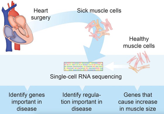
Single cell RNA sequencing facilitates the analyses of hundreds of cells and ten thousands of genes at once. The enormous amounts of data that scRNA-seq generates allow the identification of new genetic features that drive hypertrophic cardiomyopathy disease progression. Credit: Anne de Leeuw and Martijn Wehrens, copyright Hubrecht Institute.
Figure 2
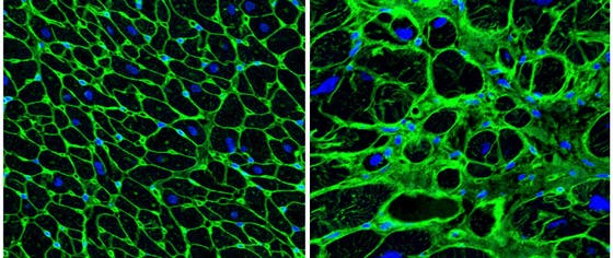
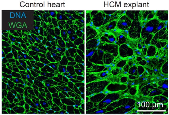
A microscopy picture of muscle cells in a normal heart (left) and muscle cells in a heart of a patient with hypertrophic cardiomyopathy (right). The black areas are single muscle cells. The muscle cells of the hypertrophic cardiomyopathy patient are bigger, and there is more space in between the cells (green), which is filled with scar tissues in the case of patients with hypertrophic cardiomyopathy. The blue indicates where the DNA of the cells can be found. Credit: Anne de Leeuw, copyright Hubrecht Institute.
Publication
Single-cell transcriptomics provides insights into hypertrophic cardiomyopathy. Martijn Wehrens*, Anne E. de Leeuw*, Maya Wright Clark*, Joep E.C. Eding*, Cornelis J. Boogerd, Bas Molenaar, Petra H. van der Kraak, Diederik W.D. Kuster, Jolanda van der Velden, Michelle Michels, Aryan Vink, and Eva van Rooij (2022). Cell Reports 2022. DOI 10.1016/j.celrep.2022.110809
*Authors contributed equally
