Stroke diagnosis, prediction and treatment monitoring with multiparametric MRI
Stroke is a major cerebrovascular disease, caused by vessel occlusion (ischemic stroke) or rupture (hemorrhagic stroke), leading to brain tissue infarction and loss of function. Treatment options are limited, and various international consortia, such as the European Stroke Network and Multi-PART, which we have joined or supported, have emphasized the need for translational image-based research to identify brain tissue that may be protected or repaired by therapeutic strategies after stroke.
Research participants
RM Dijkhuizen, WM Otte & G van Tilborg.
The availability of MRI in clinical and laboratory settings at our institute offers an ideal means for translational research in clinically relevant animal stroke models. Multiparametric information on vessel patency (MR angiography), blood flow (perfusion MRI) and tissue damage (structural MRI) can be obtained in a single session. We are applying various MRI methods to get improved insights in stroke pathophysiology, and to test novel treatment strategies in different rodent models for ischemic and hemorrhagic stroke from acute to chronic stages. Furthermore, we are developing machine learning strategies for tissue classification and outcome prediction based on multiparametric MRI data. These tools, which are also being tested in clinical settings, may help to improve diagnosis and treatment decision making.

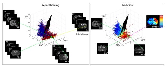
Multiparametric MRI data of brain tissue status (from maps of cerebral blood flow (CBF), apparent diffusion coefficient (ADC) and mean transit time (MTT)) in different rats with a unilateral ischemic stroke. For model training for outcome prediction, acute MRI data were voxel-wise related to tissue outcome (i.e. infarcted (red) or non-infarcted tissue (blue)) based on follow-up T2 scans or histology. Subsequently a generalized linear model (GLM)-based statistical algorithm estimated a separating plane to differentiate between infarcted and non-infarcted voxels. For tissue outcome prediction in another stroke subject, the distribution pattern of the newly introduced voxel-wise combinations relative to the separating plane can be used to estimate infarction risk (0% ≤ Pinfarct ≤ 100%). Courtesy of Mark Bouts.
